Case of the week 9.26.22
Publication Date: 2022-09-26
History
15-year-old male castrated Dachshund. Acute progression of hindlimb weakness, now paraplegic and tetraparetic, deep pain negative. Previous history of a splenic mass. Thoracic radiographs before advanced imaging.
3 images
Findings
Thorax - ventrodorsal and lateral radiographs are available for interpretation.
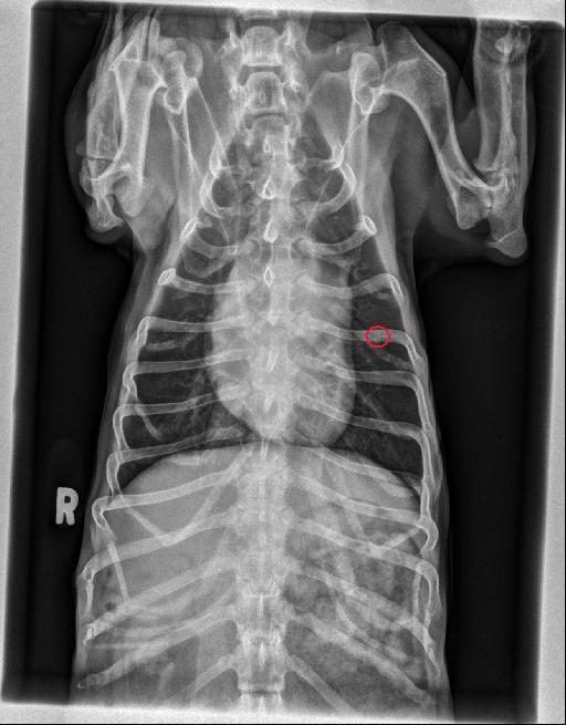
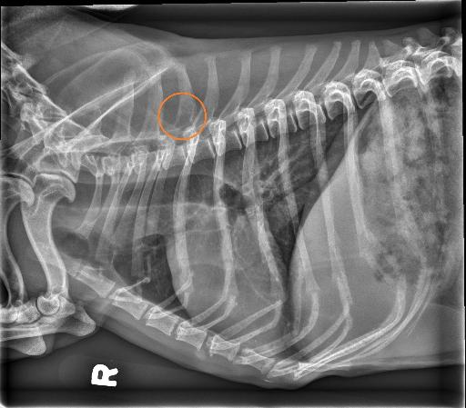
There is a mild, diffuse unstructured interstitial pulmonary pattern present which is not considered excessive given the patient’s age. There is a focal, ovoid, soft tissue opaque nodule within the caudal subsegment of the left cranial lung lobe which is superimposed with the left fifth rib. There are multiple, small, punctate to round, mineral opaque foci throughout the pulmonary parenchyma which are consistent with benign, incidental, pulmonary osteomata. The cardiac silhouette and pulmonary vessels are normal. The pleural and mediastinal spaces are normal.
There is focal moth-eaten lysis of the base of the spinous process of T3. The dorsal margin of the vertebral canal at this level is also inconspicuous. Within the included portion of the abdomen, the liver is mildly enlarged extending beyond the costal arch.
Diagnosis
- The moth-eaten lysis of the spinous process of T3 and the inconspicuous nature of the dorsal margin of the vertebral canal at this level is most concerning for a primary osseous neoplasia (such as osteosarcoma, chondrosarcoma). Metastasis from the previously removed splenic mass cannot be excluded. - The ovoid pulmonary nodule in the caudal subsegment of the left cranial lung lobe is most concerning for metastasis. - Mild hepatomegaly is non-specific and may represent benign processes (such as nodular hyperplasia) or malignancy.
Discussion
Based on those findings and the clinical signs of the patient, the owners elected for euthanasia.


Notes
This case was initially seen by Dr. Monto.
Files