Case of the week 2.7.22
Publication Date: 2022-02-07
History
14 year old female spayed Chihuahua, hematochezia
3 images
Findings
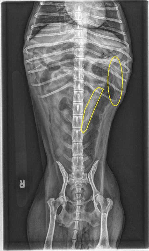
Opposite lateral and VD radiographs of the abdomen are available for interpretation.
The serosal detail is normal. The liver is small. The stomach contains a small volume of gas. The small intestines are normal and uniform in diameter. There is a moderate volume of stippled gas extending through the majority of the colonic wall, including into the pelvic canal. The colon contains a large volume of fluid and a small volume of soft tissue opaque material which has a “foamy” appearance.
The spleen, unobscured margins of the kidneys and urinary bladder are normal.
There are a few, nonspecific, small, soft tissue opaque nodules in the subcutaneous tissues of the left proximal pelvic limb.
Diagnosis
- The appearance of the colon is consistent with pneumatosis coli. Potential causes include severe bacterial colitis, colonic ulceration, or, less likely, ischemia/infarction.
Discussion
An abdominal ultrasound was performed, and underlying etiology for the pneumatosis coli was not elicited. The patient was discharged with medical treatment.

Additional references
https://radiopaedia.org/articles/pneumatosis-coli?lang=us
Files