Case of the week 2.28.22
Publication Date: 2022-02-01
History
7 year old cat. Health check up
3 images
Findings
Orthogonal radiographs of the thorax are available for interpretation.
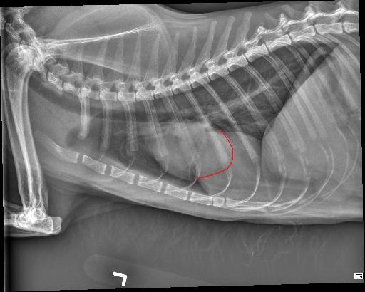
Within the caudal ventral thorax, directly adjacent to the diaphragm, there is a large, round, smoothly marginated, fat opaque structure that is causing dorsal and leftward deviation of the cardiac silhouette. The margins of the cardiac silhouette are distinctly identified superimposed with this structure on the lateral views.
The cardiac silhouette is normal in size. The pulmonary vessels, pulmonary parenchyma, and pleural space are normal.
Diagnosis
The rounded structure in the caudal ventral thorax is suspected to represent fat opaque material and may be due to herniation/eventration of falcifom fat.
Discussion
On the annotated image the cardiac margin remains visible when superimposed with the caudal structure. This indicates that those two are not of the same opacity. Furthermore the caudal structure is slightly more radiolucent compared to the cardiac outline.

Files