Case of the week 12.6.21
Publication Date: 2021-12-06
History
Bald Eagle. Found unable to fly
3 images
Findings
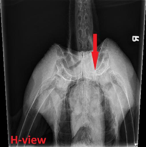
Orthogonal whole body radiographs and a caudocranial oblique radiograph of the shoulder girdle (H-view) were acquired by the emergency service. The laterality marker is incorrect on the VD and H-view.
There is a comminuted fracture of the left coracoid bone with mild displacement of the fracture fragments, best seen on the 'H' view. The margins of the fracture are mildly rounded. No new bone formation associated with the fracture is identified. There is moderate soft tissue swelling surrounding the fracture site, and the left clavicular air sac is not clearly identified.
There is a large amount of variably sized and shaped mineral opaque material in the ventriculus. No additional abnormalities of the coelomic cavity are identified.
Diagnosis
Conclusions: - Comminuted fracture of the left coracoid bone, suspected to be subacute in nature, with no evidence of osseous healing. Associated hemorrhage and/or edema and suspect collapse of the left clavicular air sac. - Mineral opacities in the ventriculus likely represent osseous fragments from a previous meal and/or incidental grit.
Discussion
https://pubmed.ncbi.nlm.nih.gov/26480013/

The patient is being treated conservatively at this point.
Files