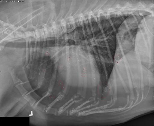
Case of the week 9.13.21
Publication Date: 2021-09-13
History
11 year old collie. Male castrated. Health check
3 images
Quizz
- Does the dog have pulmonary metastasisYes
Actually notNo
Good job
Findings
Opposite lateral and ventrodorsal views are available.
There are numerous well-defined slightly irregularly marginated mineral opaque nodules ranging in size from 1 mm to 4 mm in diameter throughout all lung lobes.
The cardiac silhouette and pulmonary vessels are normal.
There is a large amount of adipose tissue within the ventral aspect of the thorax resulting in separation of the cardiac silhouette from the sternum.
There is an incidental finding of spondylosis deformans in multiple sites of the thoracic spine, the costochondral junctions also have signs of chronic remodeling.
Diagnosis
1. Normal thorax with diffuse osteomata. There is no evidence of pulmonary metastatic disease.
Some of them are faintly circled in red to help localization without obscuring them.

Article
Here is an interesting article: https://www.vin.com/apputil/content/defaultadv1.aspx?pId=11310&id=4516254&print=1
Files