Case of the week 7.6.21
Publication Date: 2021-07-02
History
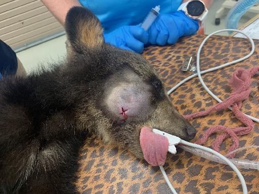
No history provided. Can you guess the species?
2 images
Findings
Protruding from the right side of the face at the level of the right zygomatic arch, there is a large soft tissue opaque swelling containing multiple gas foci. The adjacent dental structures are normal for a juvenile patient. The zygomatic arches are mildly asymmetric. There is a small step defect with discontinuous cortical margins in the caudal aspect of the right zygomatic arch. In the cranial aspect of the right zygomatic arch, there is a radiolucent line extending rostrally through the zygomatic bone close to its junction with the maxilla. This radiolucent line begins along the medial margin of the zygomatic arch and extends ~1.1cm rostrally, but does not appear to contact the lateral cortex. An ill-defined, rounded soft tissue opaque structure is seen ventral to the angles of the mandible in the lateral view. The remaining structures are normal. The patient is intubated.
Diagnosis
- The right-sided facial swelling is most consistent with an abscess, likely resulting from a previous traumatic injury. This is further supported by the suspected incomplete fractures of the right zygomatic bone, although it is possible the described lesions in the right zygomatic bone represent tangential visualization of normal suture margins. CT of the head could be considered for further evaluation if clinically indicated. The soft tissue structure ventral to the mandible is consistent with a reactive mandibular lymph node.
Discussion
The abscess was drained, and the patient was discharged to the rescue. The zygomatic arch was left in situ at this point
https://appalachianbearrescue.org/

Files