Case of the week 6.14.21
Publication Date: 2021-06-14
History
8 year old male castrate mixed breed dog. Straining to defecate
6 images
Findings
Opposite lateral and VD radiographs of the abdomen are available for interpretation.
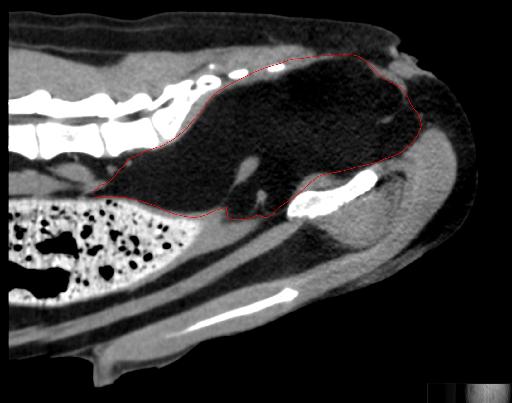
Abdomen: The serosal detail is normal. There is a broad-based, fusiform, fat to soft tissue opaque mass extending along the dorsal aspect of the pelvic canal. On the left lateral projection, the mass is fat opaque, on the right lateral projection, the mass is more soft tissue opaque. The mass is resulting in severe compression and focal narrowing of the descending colon in the pelvic canal. There is a large volume of soft tissue to faintly mineral opaque fecal material in the descending colon immediately cranial to the colonic narrowing. The stomach and small intestines are normal. The liver, spleen, unobscured margins of the kidneys and urinary bladder are normal.
Diagnosis
- Fat to soft tissue opaque mass extending along the dorsal aspect of the pelvic canal with severe focal colonic/rectal compression. This mass may be non-organ associated and located in the caudal retroperitoneal space/pelvic canal. Given its opacity, a lipoma is considered. An ill-defined or mixed density soft tissue mass (either non-organ associated or originating from the colon/rectum) is also possible.
Discussion
The patient underwent an abdominal CT for surgical planning, the CT also confirmed the fatty nature of the mass.

Notes
The case was initially seen on radiology by Dr. Johnson
Files