5.17.21
Publication Date: 2021-05-14
History
10 year Border Collie. 2 week history of melena. Lethargy.
6 images
Findings
Orthogonal radiographs of the abdomen are available for interpretation.
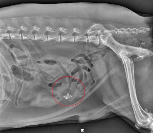
Abdomen: In the right mid-ventral abdomen, there is a soft tissue opaque mass surrounding a small intestinal segment. This small intestinal segment contains a small volume of gas and numerous, pinpoint to small mineral to metal opaque foci.
There is mild fluid streaking surrounding the mass. There is no abnormal small intestinal distention. The stomach and colon contain nonobstructive mineral opaque material but are overall normal.
The splenic tail has a mildly lobular margins with a suspect soft tissue opaque nodule extending from its ventral and dorsal margins. There is a convex soft tissue opacity extending from the ventral margins of the mid-cranial liver. The unobscured margins of the kidneys and urinary bladder are normal.
Diagnosis
Conclusions:
- Small intestinal mass with associated peritoneal effusion and/or focal peritonitis/steatitis may be of malignant (such as adenocarcinoma, lymphoma, gastrointestinal stromal cell tumor, or leiomyosarcoma) or benign (such as leiomyoma) etiology. Given the presence of a gravel sign, a chronic partial obstruction is likely. The planned abdominal ultrasound and tissue sampling are recommended for further evaluation.
- Suspect splenic nodule is nonspecific and may represent a benign or malignant process.
- Soft tissue opacity extending from the ventral margins of the liver may represent the gallbladder, however a nonspecific hepatic nodule/small mass cannot be entirely excluded.
Discussion
The patient underwent thoracic radiographs and abdominal ultrasound. On the later the mass was also visualized and there was concerned for neoplastic infiltration of the mesentery. The owner elected to not pursue any further diagnosis and the patient was euthanized

Files