Case of the week 3.29.21
Publication Date: 2021-03-26
History
2 year old Doberman, male. Cervical pain for the last 6-8months. Improves on pain medication but recurs.
4 images
Findings
Study: Multiple lateral views of the cervical, thoracic, and lumbar spine have been performed.
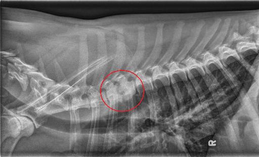
Findings: There is collapse of the T3-T4 intervertebral disc space. There is ill-defined lysis of the adjacent end-plates, with markedly irregular margination to these endplates, and there is sclerosis extending from the endplates to the mid aspect of both the T3 and T4 vertebral bodies. There is periarticular new bone formation along the dorsal and ventral margins of these endplates, giving a flaired appearance. There is minimal to mild ventral spondylosis deformans at T4-T5 and T5-T6. The remaining imaged intervertebral endplates are smoothly marginated. There is ventral spondylosis at the level of L7-S1.
Diagnosis
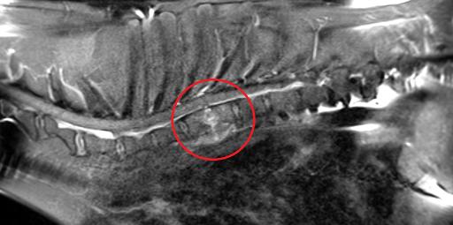
The collapse of the T3-T4 intervertebral disc space with associated endplate lysis and sclerosis, given the MRI findings, is most consistent with discospondylitis. Minimal to mild spondylosis deformans at T4-T5 and T5-T6.
Discussion
You may have noticed that there are no orthogonal radiographs for this particular study. This patient actually underwent MRI of the cervical spine initially where the discospondylitis lesion was identified. It is logistically and economically easier to evaluate the whole spine doing radiographs and allows for easier recheck rather than doing a whole body MRI.


MRI is a T1 fat sat sagittal post contrast.
Notes
Case initially seen on radiographs by Dr. Lipe
Files