Case of the week 3.8.21
Publication Date: 2021-03-08
History
9 year old Boxer. Started coughing about 3 months ago, happens 4-5 times/day
3 images
Findings
Opposite lateral and VD views of the thorax have been performed
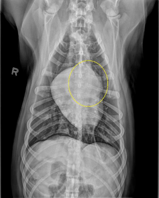
There is an ill-defined soft tissue opaque mass at the craniodorsal and left aspect of the cardiac silhouette, which is causing focal dorsal deviation of the trachea on the right lateral view, causing a “J” shape to the trachea. The remainder of the cardiac silhouette remains normal. There are a few pinpoint mineral foci within the periphery of the pulmonary parenchyma consistent with incidental osteomas, and the pulmonary parenchyma is otherwise normal. The pleural space is normal. There is multifocal incidental spondylosis deformans.
Diagnosis
Mass associated with the cardiac silhouette, consistent with a heart base mass.
Files
