Case of the week 12.14.20
Publication Date: 2020-12-14
3 images
Findings
Opposite lateral and VD radiographs of the thorax are available for interpretation.
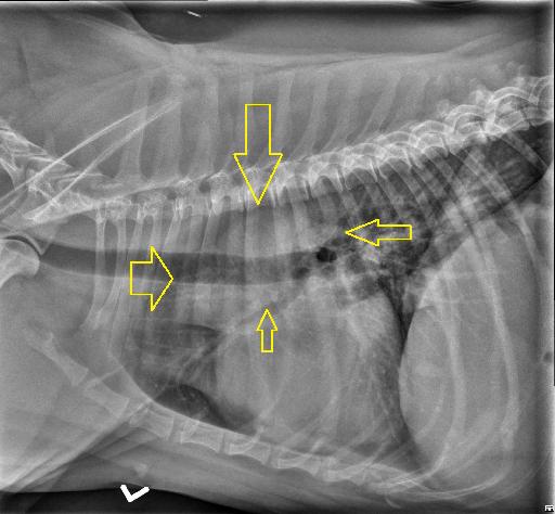
There is an ill-defined soft tissue opacity at the level of the ascending aorta, aortic arch and heart base. This opacity is resulting in mild dorsal deviation of the trachea on the right lateral projection and moderate to marked focal rightward deviation of the trachea on the VD view. There is a mild, diffuse, unstructured interstitial pattern, not considered excessive for the patient’s age. The pulmonary vessels are normal. There is a fusiform soft tissue nodule superimposed with the right caudal lung lobe and liver at the level of the 7th intercostal space, only seen on the left lateral projection. A small soft tissue nodule is superimposed with the subcutaneous tissues of the cranioventral thorax.
Diagnosis
- The described soft tissue opacity is most consistent with a heart base tumor, such as a chemodectoma. An echocardiogram or thoracic CT can be performed for further evaluation.
- Fusiform soft tissue nodule superimposed with the right caudal lung lobe likely represents superimposition of structures (such as a cutaneous/subcutaneous nodule). Cutaneous nodules are nonspecific and may be of benign or malignant etiology.
Discussion
The patient was confirmed on CT and echocardiography to have a heart base mass, most consistent with a chemodectoma.

Files