Case of the week 11.9.20
Publication Date: 2020-11-06
History
14 year old domestic shorthair cat, losing weight for 3-4 weeks
3 images
Findings
Left lateral, right lateral, and ventrodorsal abdominal radiographs are available for review.
A round to lobulated soft tissue opaque mass measuring at least 7 cm in greatest diameter is seen in the mid abdomen, circumferentially displacing the intestinal tract. The small intestines contain gas and heterogeneous soft tissue opaque material. No overt intestinal distension is identified. The stomach contains a small amount of gas and the colon is gas filled. There is wispy soft tissue opacity throughout the ventral abdomen. The liver, spleen, visible margins of the kidneys, retroperitoneal space, and urinary bladder are normal. There is nominal incidental spondylosis deformans at the lumbosacral space. The caudal thorax is normal.
Diagnosis
- Lobulated mid-abdominal mass may be originating from the gastrointestinal tract, mesentery, or represent multiple enlarged mesenteric lymph nodes. A malignant neoplastic process, such as lymphoma or other round cell neoplasia, is considered most likely. The suspected abdominal effusion may be a non-specific transudate, neoplastic exudate, and/or hemorrhagic effusion.
Discussion
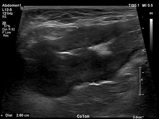
On ultrasound the following was noted: Transmural, circumferential mass arising from the wall of the ascending/transverse colon extending to the level of the ileocolic junction is most consistent with malignant colonic neoplasia, such as lymphoma, especially given the concurrent marked colic lymphadenopathy. Other differentials including leiomyosarcoma/GIST, or adenocarcinoma cannot be excluded. Marked colic lymphadenopathy is most consistent with metastatic/disseminated disease. Hypoechoic tendril-like extension from the mass and colic lymph node is most consistent with lymphomatosis/carcinomatosis/sarcomatosis

The cat was diagnosed with lymphoma based on FNAs
Files