Case of the week 11.2.20
Publication Date: 2020-10-28
History
6 month old Australian Shepherd. Ran into a couch. Intermittent ataxia
2 images
Findings
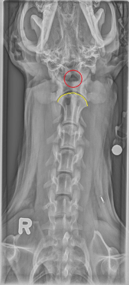
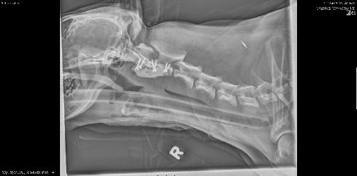
Right lateral and VD radiographs of the neck are available for interpretation.
A normal dens of C2 is not identified on any of the radiographs. On the straight lateral projection, there is the impression of the ventral aspect of the C2 spinous process being in a slightly ventral location with respect to the dorsal aspect of C1. An ill-defined, rounded to ovoid, faint osseous structure is identified cranial to the body of C1 on the VD projection. There are open physes, consistent with the patient’s young age.
Diagnosis
Absence of a normal dens with concern for atlantoaxial instability, given the mild ventral displacement of C2 in relation to C1. Osseous fragment cranial to C1 may represent the fractured or avulsed dens.
Discussion
The patient underwent both CT and MRI which confirmed the AA subluxation. The dens axis was confirmed fracture. The patient was treated conservatively while waiting for 3D printed guides. Surgery was performed at the end of last week.


Notes
The case was initially seen by Dr Debow and Fazio
Files