Case of the week 9.8.20
Publication Date: 2020-09-04
3 images
Findings
Abdomen: Opposite lateral and VD projections of the abdomen are available for interpretation.
The patient is markedly thin, seen by tenting of the skin over the spinous processes of the thoracic and lumbar vertebrae. The serosal margin detail is diffusely decreased.
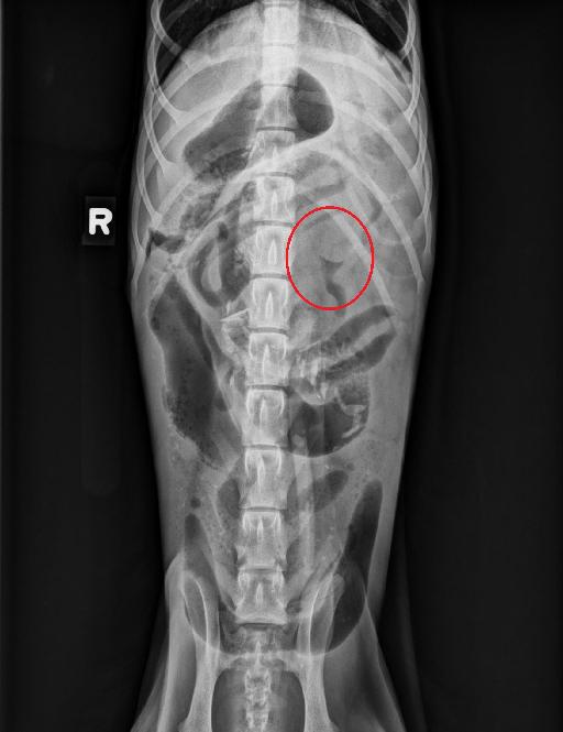
The stomach contains a small amount of gas and granular material and is minimally distended. Numerous small intestinal segments are markedly gas distended while other small intestinal segments are normal in diameter and are fluid-filled or empty. Mixed soft tissue and gas opaque granular material as well as numerous variably sized and shaped mineral opacities are also seen throughout the gastrointestinal tract, some within the colon and some within small intestinal segments. A well-defined, convex soft tissue opacity is seen on the VD projection, to the left of the L1 vertebra, which is outlined by gas within the adjacent intestinal segment.
The liver is normal in size with sharp caudal ventral margins. The spleen is not clearly seen. The kidneys are mostly obscured on the VD projection however the visible portion of the kidneys on the lateral views are normal. The urinary bladder is normal.
The included portion of the thorax is normal. The visible musculoskeletal structures are normal.
Diagnosis
Conclusion:
- Gastrointestinal changes are consistent with mechanical ileus, most likely secondary to intussusception. Numerous mineral opacities throughout the gastrointestinal tract are consistent with ingestion of foreign material such as bone. Surgery is recommended. Abdominal ultrasound could be performed prior to surgery to further assess the gastrointestinal tract.
- Decreased serosal margin detail is most likely due to the thin body condition of the patient, however a small amount of peritoneal effusion cannot be entirely ruled out.
Discussion
The patient underwent abdominal surgery and the intussusception was confirmed. This was reduced and the patient is doing fine.

Notes
This case was initially seen by Dr. Auger
Files