Case of the week 8.31.20
Publication Date: 2020-08-28
3 images
Findings
Orthogonal radiographs of the thorax are available for interpretation.
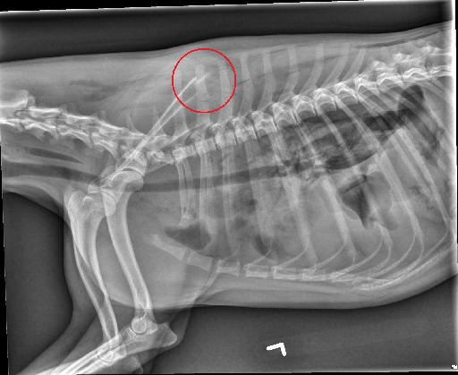
There is a moderate volume of pleural effusion bilaterally, more severe in the right hemithorax. There are multiple, variably-sized gas foci in the pleural space bilaterally. There is retraction of the caudal lung lobe margins from the thoracic wall.
There is a moderate to severe multifocal unstructured interstitial pattern bilaterally, most severe in the right middle lung lobe. The caudal vena cava is small. The unobscured margins of cardiac silhouette are normal.
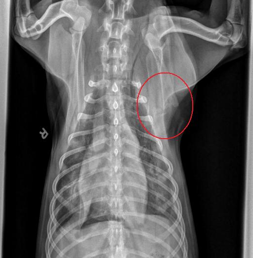
There are minimally displaced fractures of the proximal aspect of the left 2nd and 4th ribs and suspected minimally displaced fracture of the left 1st rib.
There is a segmental fracture of the proximal left third rib resulting in a separate small fragment which is mildly cranially displaced. There is mild subcutaneous emphysema of the cervical soft tissues and extending along the dorsum of the thorax and abdomen.
Diagnosis
Did you notice the extra finding ?
- Pleural effusion (consistent with the reported hemothorax), bilateral pneumothorax, multiple left rib fractures, LEFT SCAPULAR FRACTURE and subcutaneous emphysema are consistent with the reported trauma. The pulmonary pattern likely represents a combination of pulmonary contusions and atelectasis.
- Evidence of hypovolemia/shock.
- Unremarkable abdomen.


Files