Case of the week 8.24.20
Publication Date: 2020-08-24
3 images
Findings
Orthogonal radiographs of the abdomen are available for interpretation.
A smoothly marginated, oval-shaped soft tissue mass measuring 6.1cm x 3.7cm abuts the dorsal mid to caudal margin of the splenic tail. A large amount of mineralized material is present in the stomach and colon with smaller amounts in the small intestines. The small intestines are otherwise empty and normal in diameter. The liver, visible portions of the kidneys, retroperitoneal space, urinary bladder, and serosal detail are normal.
Metal opaque suture material is present in the locations of the ovariohysterectomy ligatures. The imaged skeletal structures are normal.
Diagnosis
- Cranioventral abdominal mass is likely arising from the splenic tail (lymphoid hyperplasia, hematoma, hemangiosarcoma, other sarcoma), although an eccentric intestinal mass or mesenteric mass cannot be completely ruled out. - Large amount of ingested foreign mineral material; no evidence of mechanical obstruction.
Discussion
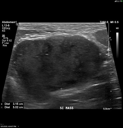
The dog underwent thoracic radiographs which showed no evidence of pulmonary metastasis. On abdominal ultrasound the mass was seen arising from the small intestines. The mass was resected surgically and was consistent on histopathology as either a leiomyosarcoma or a gastrointestinal stromal tumor (GIST).

Notes
The case was initially seen on radiology by Dr. Morandi
Files