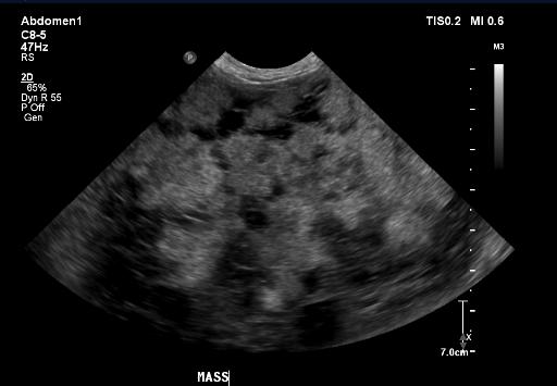
Case of the week 8.17.20
Publication Date: 2020-08-13
History
11 year old mixed breed dog. Abdominal distention.
3 images
Findings
Orthogonal radiographs of the abdomen are available for interpretation.
The serosal detail is normal.
Within the left mid to caudal dorsal abdomen, there is a large (at least 13L x 9.6W x 1.0H cm), smoothly marginated, ovoid, soft tissue opaque mass which is causing rightward and ventral deviation of the small intestines and colon. A normal left kidney is not identified. There are a few punctate to linear mineral foci within the right kidney, which measures within normal limits. The liver, spleen, GI tract, and urinary bladder are normal. There is mild incidental spondylosis deformans.
Diagnosis
The large abdominal mass is most consistent with a severely enlarged left kidney, which may be secondary to renal neoplasia (such as carcinoma) or to severe hydronephrosis. An abdominal ultrasound is recommended for further evaluation.
Discussion
The patient underwent an abdominal ultrasound and thoracic radiographs. The mass was subsequently removed and was diagnosed as a transitional cell carcinoma. The patient is still doing well 18 months post-surgery.

Notes
This case was initially seen by Dr. Auger.
Files