Case of the week 8.3.20
Publication Date: 2020-08-03
3 images
Diagnosis
There is a large, mineral opaque structure in the caudal esophagus, resembling the shape of a portion of a lumbar vertebra [measuring up to 3.9 (H) x 5.1 (L) cm]. The esophagus surrounding this structure is moderately focally distended and contains a small volume of fluid and gas at this level. There are a few, small gas foci in the plane of the cranial vena cava and the right ventricle. The cardiovascular structures are otherwise normal. There is an unstructured interstitial pattern, not considered excessive for the patient’s age. There are a few, punctate, mineral opaque foci in the periphery of the pulmonary parenchyma, consistent with benign pulmonary osteomas.
Discussion
Caudal esophageal foreign body. Endoscopic retrieval is recommended. No definitive of pneumomediastinum.
Notes
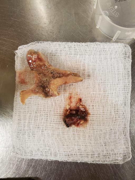
A T-bone was removed by endoscopy. The patient is doing well.
Thanks to Drs. Olin and Ryan for the photograph

Files