Case of the week 7.13.20
Publication Date: 2020-07-12
History
9 year old cat. Waxing and waning diarrhea for the last 5 months
3 images
Findings
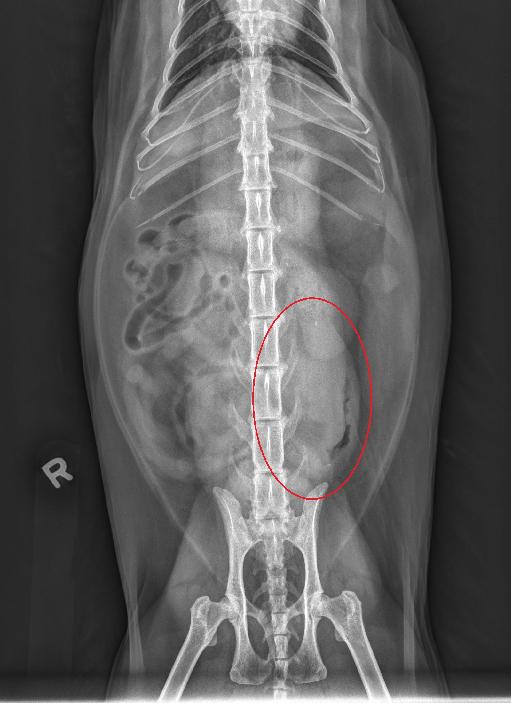
Opposite lateral and VD radiographs of the abdomen.
The serosal detail is normal. The liver and spleen are normal. The stomach is relatively empty with only a very small amount of gas in it.
The small intestines are diffusely normal in diameter with a few incidentally having a string of pearls pattern. On the VD view, in the left caudal abdomen, there is an ill-defined ovoid region of increased soft tissue opacity and mass effect superimposed with the margins of the left kidney caudal pole, and border effaces with a segment of small intestine in the left caudal lateral abdomen.
On the lateral views, immediately ventral to the left kidney, and superimposed with a string of pearls gas dilated intestinal segment, there is an ill-defined, fusiform-shaped soft tissue opacity.
There is an ill-defined rounded area of increased soft tissue opacity at the medial aspect of the splenic tail on the VD view. The kidneys are mildly irregularly marginated, and have a few pinpoint ill-defined mineral foci in the plane of their pelvises. The urinary bladder is normal.
The included thoracic and musculoskeletal structures are normal.
Diagnosis
Left caudal lateral, ill-defined, abdominal mass, likely arising from the large intestinal tract. Suspect concurrent mesenteric lymphadenopathy. Consideration is given to neoplasia, such as lymphoma or adenocarcinoma.
The ill-defined round soft tissue opacity medial to the splenic tail likely represents either soft tissue summation of the left pancreatic limb or nodular hyperplasia of the pancreas.
Discussion
The patient underwent an abdominal ultrasound which confirmed the presence of a colonic mass with marked colic lymphadenopathy.
No changes associated with the pancreas were identified.
On FNAs of the colic lymph node and colonic mass the patient was diagnosed with large cell lymphoma.

Files