Case of the week 6.8.20
Publication Date: 2020-06-08
History
6 year old Boxer, coughing for 6 months.
3 images
Findings
Orthogonal radiographs of the thorax.
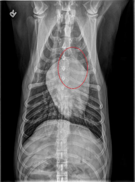
There is an ill-defined, increased, soft tissue opacity of the cranial mediastinum (better appreciated on the lateral views) which subtly protrudes ventrally. On the VD views, there is mild rightward deviation of the caudal intrathoracic trachea.
There is a diffuse mild to moderate unstructured interstitial pattern. The cardiovascular structures are normal. The remaining thoracic structures are normal.
Diagnosis
- Ill-defined cranial mediastinal mass may represent a neoplastic process, such as lymphoma or thymoma or ectopic thyroid carcinoma; or less likely a reactive cranial mediastinal lymphadenopathy of infectious or inflammatory etiology. The rightward deviation of the trachea may be due to mass effect or normal variation.
- The pulmonary pattern is nonspecific and is likely due to age-related changes and/or underinflation; however, an infiltrative process (such as lymphoma) cannot entirely be ruled out.
Discussion
Ultrasound guided FNAs of the mass were performed. The results came back as epithelial tumor such as chemodectoma.

Files