Case of the week 4.27.20
Publication Date: 2020-04-27
6 images
Findings
Orthogonal radiographs of the abdomen are available for interpretation.
The serosal detail is normal.
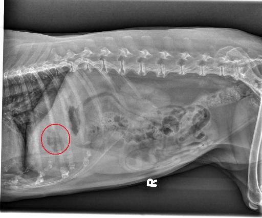
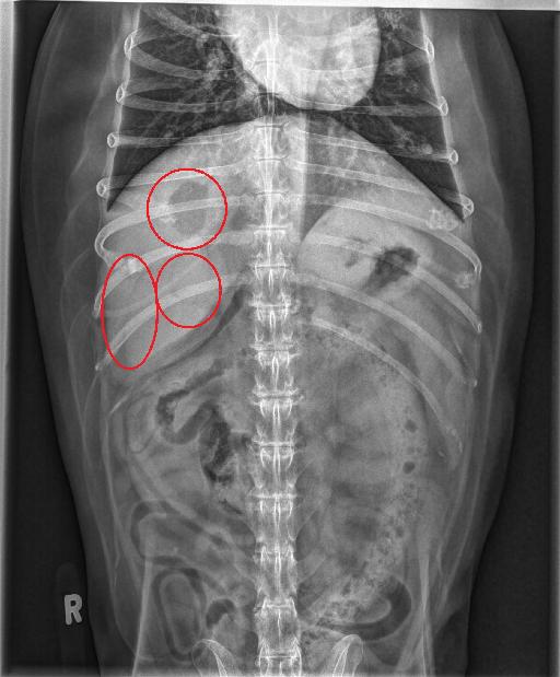
The liver is moderately enlarged extending beyond the costal arches with rounded caudal margins. There is a large, irregularly marginated and rounded gas focus superimposed with the right cranial aspect of the liver, in the region of the gallbladder and a few, adjacent, smaller gas foci. There are linear fat to gas opaque foci superimposed with the right liver.
The spleen, kidneys, urinary bladder and gastrointestinal tract are normal.
There are multifocal thoracolumbar spondylosis deformans. There is narrowing of the T12-T13 intervertebral disc space with sclerosis of the endplate. There is moderate to severe coxofemoral periarticular new bone formation.
Diagnosis
- Gas focus in the region of the gallbladder is most consistent with emphysematous cholecystitis although a hepatic abscess or a necrotic hepatic neoplasm cannot be excluded. Linear fat to gas opaque foci superimposed with the right liver may represent gas within the intrahepatic ducts or may be fat dissecting between liver lobes. Static hepatomegaly is nonspecific and may represent acute hepatitis or may be secondary to other benign or malignant processes.
- T12-T13 degenerative intervertebral disc disease.
Annotated radiographs and notes


Case initially seen by Dr. Auger.
Files