Case of the week 3.30.20
Publication Date: 2020-03-30
History
13 year old female spayed dog. History of anal gland adenocarcinoma 2 years aago (AGASACA)
6 images
Findings
Orthogonal radiographs of the abdomen are available for intepretation
The serosal detail is within normal limits.
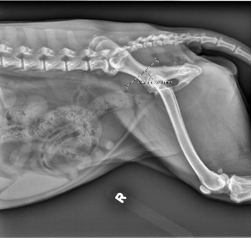
There is a well defined, ovoid, soft tissue opaque mass, measuring approximately 6.5cm in diameter, within the dorsal aspect of the pelvic canal causing marked ventral deviation of the colon.
The liver is mildly diffusely enlarged, extending past the costal arch. The spleen is mildly enlarged. The kidneys and urinary bladder remain normal. The GI tract is otherwise normal.
The GI tract is otherwise normal. There remains incidental lumbosacral spondylosis deformans.
Diagnosis
- Large pelvic mass, most consistent with a metastatic lymph node (sacral vs internal iliac) from the historic apocrine gland anal sac adenocarcinoma (AGASACA). A non-lymph node associated mass (metastatic or primary) is considered less likely.
- Hepatomegaly and splenomegaly is non-specific and likely represent benign etiologies. Malignant etiologies are considered less likely.

Notes
Case initially seen by Dr Morandi
Files