Case of the week 3.23.20
Publication Date: 2020-03-23
History
9 year old mixed breed dog, male castrated. 6 week history of progressive left hind limb lameness.
3 images
Findings
Orthogonal radiographs of the abdomen are available for interpretation.
The serosal detail is normal.
The prostate is enlarged and lobular, with multiple pinpoint mineral foci within its parenchyma. The prostate occupies approximately 40% of the height of the pelvic inlet.
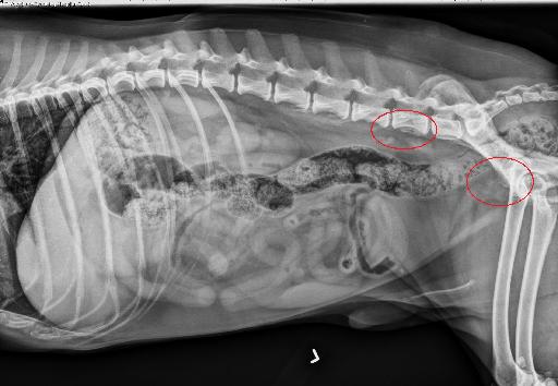
There is an ill-defined, ovoid soft tissue opacity just ventral to L6-L7, causing mild ventral deviation of the colon. Best seen on the left lateral view just ventral to the L5-L6 intervertebral disc space, there are 2 round soft tissue to mineral opaque structures.
There is irregular periosteal proliferation along the ventral aspect of the vertebral bodies of L6 and L7. There is similar irregular periosteal proliferation along the lateral aspect of the body of the left ilium, just cranial to the acetabulum, with mild sclerosis of the surrounding bone, and punctate lytic lesions.
The liver, GI tract, and spleen are normal. The lateral margin of the left kidney is mildly flattened at its cranial aspect. There is a round soft tissue nodule within the subcutaneous tissues dorsal to the caudal lumbar spine.
Diagnosis
The appearance of the prostate, with pinpoint mineralizations, is most consistent with a neoplastic process (prostatic carcinoma) associated sublumbar lymphadenopathy, which is most consistent with metastatic disease. These described osseous lesions of L6, L7, and the ilium are consistent with osseous metastasis.

Article
This is an excellent article to review: https://www.ncbi.nlm.nih.gov/pubmed/19400462
If you're short on time the take home message is: Neutered dogs with prostatic mineralization were very likely to have prostatic neoplasia (PPV 100%). Intact dogs were unlikely to have prostatic neoplasia if no mineralization was found on radiographs or ultrasound.
Notes
Case initially seen by Dr. Lipe
Files