Case of the week 3.9.20
Publication Date: 2020-03-08
3 images
Findings
Orthogonal radiographs of the pelvis are available for interpretation.
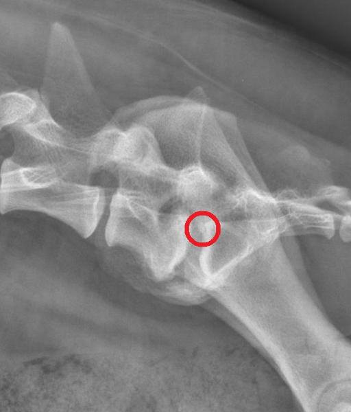
There is a moderate sclerosis at the caudal endplate of L7 and cranial endplate of S1, with remodeling of the endplate. There is moderate ventral spondylosis noted. At the dorsal aspect of the cranial endplate of S1, there is a small well defined round osseous fragment.
Overall the coxofemoral joints are considered to be within normal limits.
Incidentally there is a metallic suture superimposed with the dorsal aspect of the urinary bladder likely secondary to previous neutering.
Diagnosis
Impression: Changes most suggestive of OCD of the cranial endplate of S1. Secondary remodeling changes of L7-S1. If clinically indicated and for further evaluation advanced imaging could be considered.

Literature
For those interested here is an article discussing this pathology: https://doi.org/10.1111/j.1751-0813.2009.00418.x
Notes
Case initially seen by Dr Hespel
Files