Case of the week 2/3/20
Publication Date: 2020-02-03
History
12 year old mixed breed dog, inapettence, lethargy
3 images
Findings
There is decreased serosal detail, most severe in the mid-ventral abdomen.
The liver is moderately enlarged extending beyond the costal arches with lobular margins and is causing caudodorsal deviation of the gastric axis.
There is an ill-defined, rounded to lobular soft tissue opaque structure in the mid-ventral abdomen caudo-dorsal to the liver. There are intestinal segments superimposed with this structure on all views. The cranial pole of the left kidney is rounded and enlarged compared to the caudal pole. The unobscured margins of the right kidney are normal. The spleen and urinary bladder are normal. No free peritoneal gas is identified. T
here is incidental spondylosis deformans at T11-12, T12-13. The included caudal thorax is normal.
Diagnosis
- Abdominal mass may be intestinal or lymph node in origin. Moderate hepatomegaly, soft tissue mass and mild peritoneal effusion raises concern for a diffuse infiltrative neoplasia (lymphomatosis or carcinomatosis). Abdominal ultrasound is recommended for further evaluation.
- The appearance of the left kidney may represent a renal mass/nodule cannot be entirely excluded.
Discussion

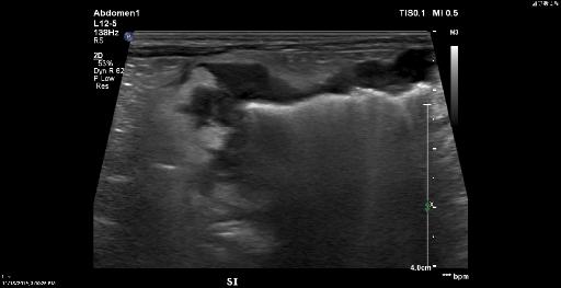
The patient underwent abdominal ultrasound where the following was found.
Jejunal wall mass with regions of ulceration, multifocal sites of asymmetric jejunal and colonic wall thickening (with a focal region of loss of colonic wall layering), bilateral renal nodules/small masses, and nodular hepatomegaly with hepatic nodules/small masses; these findings are most consistent with disseminated neoplasia, with primary consideration given to lymphoma.
Files