Case of the week 1.13.2020
Publication Date: 2020-01-13
History
10 year old Shih Tzu, Large mass on the right side of the neck. FNAs came back as severe hemorrhage.
3 images
Findings
Opposite lateral and VD radiographs of the thorax are available for interpretation.
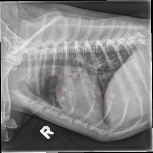
There are multiple, round, ill-defined, soft tissue opaque nodules throughout the pulmonary parenchyma (measuring up to approximately 6.5 mm). The cardiovascular structures, mediastinal structures and pleural space are normal. The included cranial abdomen is normal.
The T13 vertebra is transitional with a hypoplastic left rib.
Diagnosis
- Multiple pulmonary nodules are concerning for pulmonary metastatic disease secondary to the reported cervical mass.
- Incidental transitional T13 vertebra.
Discussion
The owner declined further diagnosis upon discovery of the pulmonary metastasis. It is likely considering the location of the mass and its hemorrhagic nature that it represents a neoplastic process arising from the thryoid such as a thyroid carcinoma.

This case remains classic in its appearance of pulmonary metastasis.
Files