Case of the week 12.30.19
Publication Date: 2019-12-30
History
3 year old female Chihuahua. Hemorrhagic gastroenteritis
3 images
Findings
Opposite lateral and VD radiographs of the abdomen are available for interpretation.
The serosal detail is normal.
There is a large, round, well-defined, soft tissue opaque structure in the mid-ventral abdomen causing cranial displacement of the intestinal tract and dorsal displacement of the colon.
The liver, head of the spleen, visible margins of the kidneys, and gastrointestinal tract are normal.
Diagnosis
Given the intact status of the patient, enlarged mammae and vulva, and the location, the large structure in the mid-abdomen is most likely originating from the reproductive tract (i.e. uterus).
Differential diagnosis include a mid-gestational pregnancy, a fluid-filled uterus (such as pyometra, mucometra, hydrometra), or less likely a uterine mass. A splenic mass is considered unlikely given its location.
An abdominal ultrasound is recommended for further evaluation.
Discussion
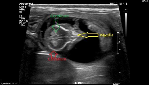
An abdominal ultrasound was performed. There were changes consistent with colitis and enteritis. Mild abdominal reactive lymphadenopathy. Additionally there was one single viable fetus.

Files