12.23.19
Publication Date: 2019-12-23
History
12 year old Chihuahua, history of heart disease, increased expiratory noise.
3 images
Findings
Opposite lateral and VD projections of the thorax are available for interpretation.
There is underinflation of the lungs on all lateral views.
A small focal bulge is noted at the caudal dorsal aspect of the cardiac silhouette on the left lateral views which is not corroborated on the right lateral views. The cardiac silhouette is otherwise normal in overall size and the pulmonary vasculature is normal.
There is a diffuse moderate bronchial and unstructured interstitial pattern.
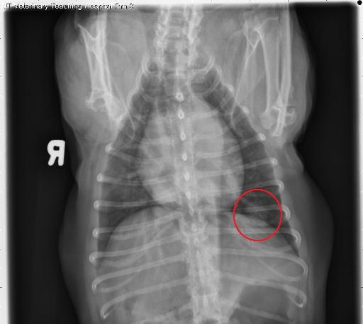
Best seen on the left lateral views, a round, ill-defined, approximately 1.4 cm, soft tissue opaque nodule is noted in the plane of the ventral aspect of the right caudal lung lobe, in the sixth intercostal space, superimposed with the diaphragm and cardiac silhouette.
There is the impression of a round region of radiolucency superimposed with this nodule. This nodule is not corroborated on the VD or DV views.
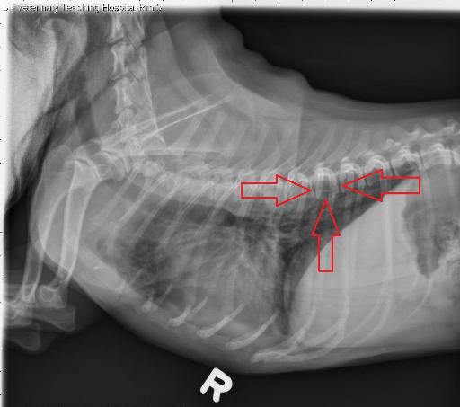
There is the impression of a broad-based, ovoid, soft tissue opaque nodule ventral to the T6 and T7 vertebrae on the right lateral and left lateral views, superimposed with the descending aorta. This possibly corresponds to an ill-defined, rounded soft tissue opaque nodule in the plane of the left caudal lung lobe on the DV view, superimposed with the left eighth rib.
On all lateral views, there is the impression of cortical discontinuity of the ventral aspect of the T8 vertebral body as well as the impression of moth-eaten lysis of the T8 vertebral body. There is also the impression of ill-defined periosteal proliferation along the ventral aspect of the T8 vertebral body. These findings are not clearly corroborated on the VD or DV views.
Diagnosis
- At least two suspected pulmonary nodules (right caudal and left caudal lung lobes), one of which appears cavitary, are concerning for pulmonary metastatic disease.
- Aggressive lytic and proliferative changes to the T8 vertebra may represent metastatic disease or to primary bone neoplasia (such as osteosarcoma).
- Equivocal left atrial enlargement may represent a patient variant or may be secondary to chronic degenerative mitral valve disease. There is no radiographic evidence of cardiac decompensation.
Files

