11.18.19
Publication Date: 2019-11-18
History
12 year old labrador retriever. mass over the left hip
3 images
Findings
Opposite lateral and VD views of the thorax have been performed.
The cardiovascular structures are normal.
There are multiple soft tissue opaque nodules throughout the pulmonary parenchyma, measuring up to 0.7 cm in diameter.
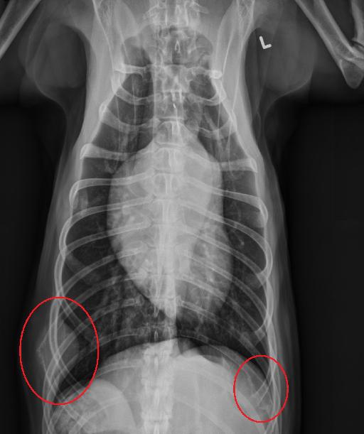
There is a broad based soft tissue opaque mass along the lateral aspect of the right body wall and extending into the pleural space, centered at the body of the right 9th rib. There is focal motheaten lysis of this rib with spiculated periosteal reaction, and a pathologic fracture. There is an expansile lesion with motheaten lysis in the dorsal body of the left 11th rib, with irregular periosteal proliferation. There is a broad based, subcutaneous, fat opaque structure along the ventral body wall, at the level of the 4th and 5th sternebrae consistent with an incidental lipoma. This structure is superimposed with the costochondral junction of the left 5th rib on the VD view.
Diagnosis
The pulmonary nodules, aggressive rib lesions, and changes to the spleen are most consistent with metastatic disease.
Discussion
The patient was confirmed to have a lymphangiosarcoma.
Lymphangiosarcoma is a rare tumor that arises from the lymphatic endothelial tissue (cells that line lymphatic vessels). Unfortunately this tumor type carries a medium to high metastatic potential.

Files