10.21.19
Publication Date: 2019-10-21
History
9 year old Australian cattle dog, acute blindness, acute vomiting, hypoglycemic, signs of sepsis
4 images
Findings
Orthogonal radiographs of the thorax are available for interpretation.
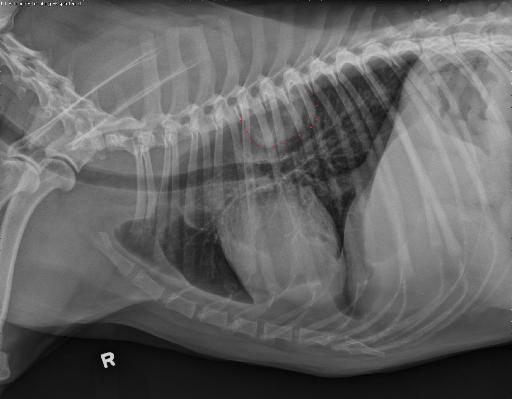
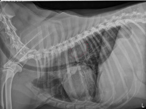
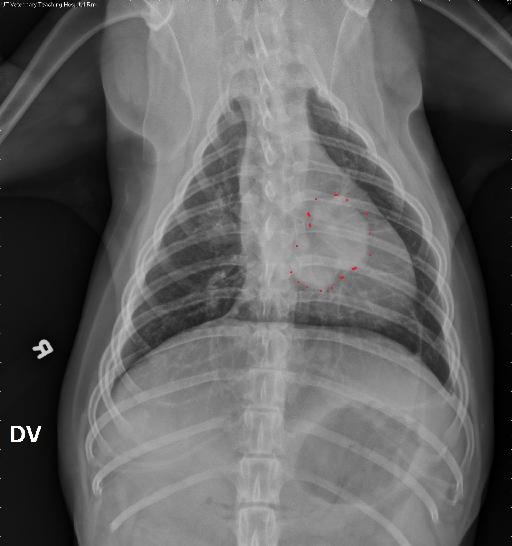
Within the left dorsal thorax, there is a rounded and lobular soft tissue mass, extending from left ribs 6 through 9. This mass appears broad-based on the right lateral projection.
The cardiovascular structures are normal.
There is a small amount of pleural effusion, causing thin pleural fissure lines.
There is a mild diffuse bronchial and unstructured interstitial pattern, not considered excessive for the patient’s age.
Diagnosis
The described mass in the left dorsal thorax (in addition to the described pleural effusion) may originate from the thoracic wall and represent a neoplastic mass (such as chondrosarcoma), or an abscess. A pulmonary mass (such as carcinoma) is also considered.
Discussion and annotated images
Note how much more conspicuous the mass is on the DV vs VD.
The patient concurrently had abdominal effusion and mild hepatomegaly. The owners elected for euthanasia and on necropsy was diagnosed with highly disseminated aggressive histiocytic sarcoma with widespread involvement of major organ systems, including the brain.


Files