9.30.19
Publication Date: 2019-09-30
History
8 year old Cane Corso. Lameness
3 images
What did you see ?
Did you see that on those radiographs ?


More images


Findings
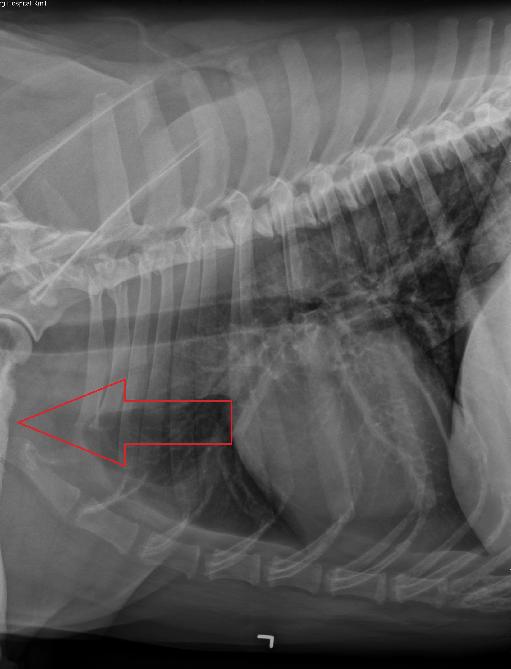
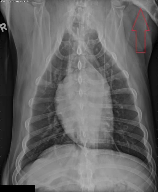
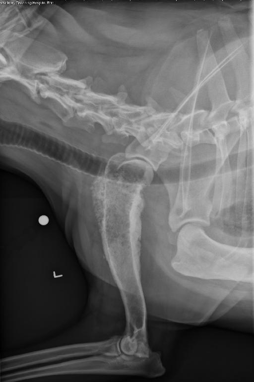
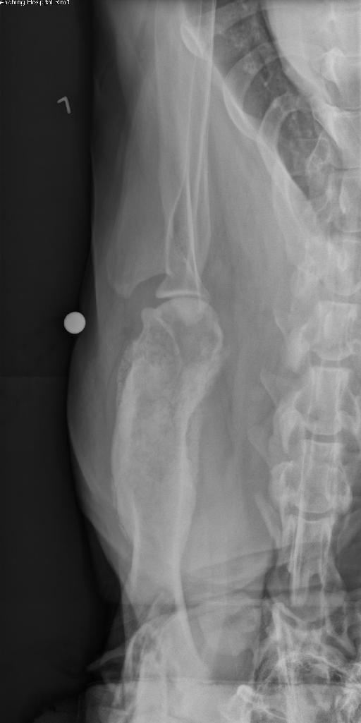
Opposite lateral and VD views of the thorax, and orthogonal views of the left humerus have been performed.
The cardiovascular structures are normal. There is a mild diffuse unstructured interstitial and bronchial pattern, not considered excessive for the patient’s age. The pleural space and mediastinum are normal. There is multifocal incidental spondylosis deformans, and periarticular new bone formation surrounding the caudal thoracic articular facets, consistent with chronic degenerative changes.
There is a lytic and proliferative osseous lesion affecting the proximal metaphysis and proximal half of the diaphysis of the left humerus. There is moth-eaten permeative lysis of the proximal humerus, with a long transition zone. There is columnar periosteal proliferation circumferentially around the proximal humerus. There is thinning of the cortex of the proximal humerus, most severe at its cranial and medial aspect. There is mild elbow degenerative joint disease.
Diagnosis
The described lytic and proliferative lesion affecting the proximal left humerus is most consistent with primary osseous neoplasia (such as osteosarcoma). Fungal osteomyelitis of (such as blastomycosis) cannot be entirely excluded.
- Normal geriatric thorax, without evidence of pulmonary metastatic disease.
Diagnosis
The humeral lesion was confirmed to be an osteosarcoma. The patient is undergoing treatment currently.