8.19.19
Publication Date: 2019-08-19
History
10 month old female spayed mixed breed dog. Multiple episodes of vomiting and diarrhea for 2 days.
6 images
Findings
Orthogonal radiographs of the abdomen are available for interpretation.
Serosal detail is normal.
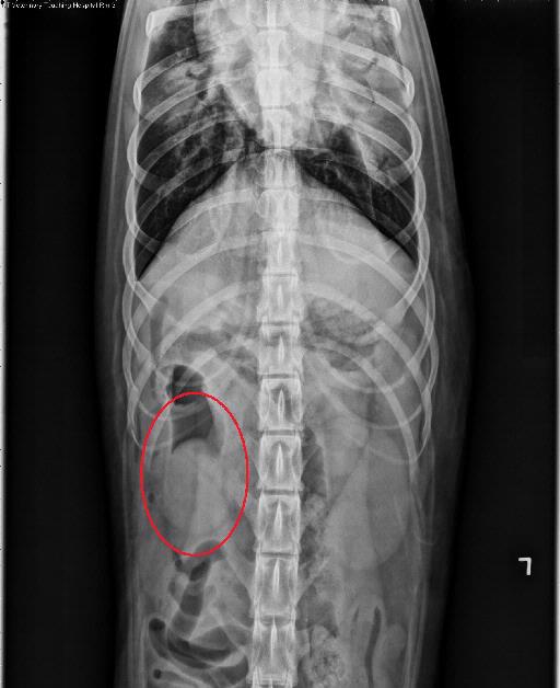
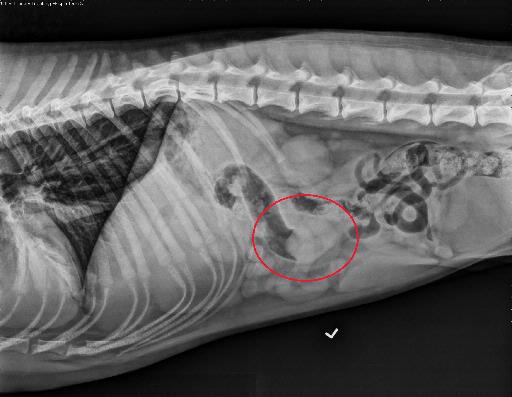
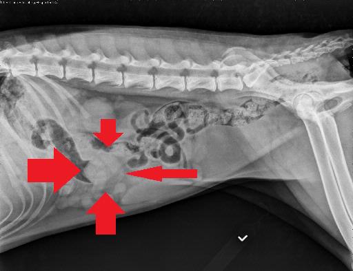
The stomach and small intestines are normal. Seen on the left lateral and VD views, there is a convex soft tissue opaque structure extending into the lumen of the ascending colon, which is gas dilated.
The liver, spleen, kidneys, and urinary bladder are normal. There are open physes consistent with the young age of the patient.
There is a patchy unstructured interstitial to alveolar pattern within the ventral aspects of the right middle, and caudal subsegment of the left cranial lung lobes
Diagnosis
The described convex soft tissue opaque structure within the ascending colon is most concerning for an ileocolic intussusception.
The pulmonary changes are consistent with severe aspiration pneumonia, secondary to the reported vomiting.
Discussion
An intussusception was confirmed on ultrasound. The patient underwent surgery and recovered well.
On the labelled radiographs the intussusception is circled in red


Files