Case of the week 8.5.19
Publication Date: 2019-08-05
History
9 year old bengal cat. 3-4 days of hyporexia, lethargy, currently febrile
3 images
Findings
Orthogonal radiographs of the abdomen are available for interpretation.
The serosal detail in the mid abdomen is decreased.
There is an ill-defined, round, soft tissue opaque mass within the mid abdomen. This mass is causing dorsal and ventral deviation of the small intestines (mass effect). The liver is mildly enlarged, extending past the costal arch, and causing caudal deviation of the gastric axis. The stomach contains a small amount of gas.
There is a mildly dilated small intestinal segment within the cranial ventral abdomen, which is more dilated than the remaining small intestines. The colon is normal. The spleen is markedly diffusely enlarged, extending caudally to the level of the urinary bladder with smooth margins. The kidneys are mildly irregularly marginated, and measure within normal limits. The urinary bladder is normal.
Diagnosis
The mass in the mid abdomen likely represents a severely enlarged mesenteric lymph node. An intestinal mass cannot be entirely excluded. The described dilated small intestinal segment is likely transient, however, dilation orad to an intestinal mass cannot be entirely excluded. Given the concurrent splenomegaly and hepatomegaly, lymphoma is considered most likely. An abdominal ultrasound is recommended for further evaluation.
Discussion
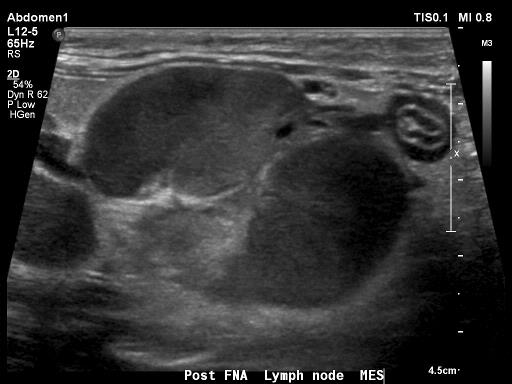
Ultrasound of the abdomen was performed, and FNAs of the identified enlarged and abnormal mesenteric lymph node as well as of the spleen were both conclusive for large cell lymphoma.

Files