Case of the week 7.29.19
Publication Date: 2019-07-29
History
7 year old English bulldog. Anemia
3 images
Findings
Opposite lateral and VD radiographs of the abdomen.
There is generalized decreased serosal detail characterized by fluid/soft tissue opaque streaking and visceral crowding.
The spleen is diffusely moderately to markedly enlarged. The splenic head is not clearly seen on the VD view. There is a mass effect of the left lateral and mid abdomen, resulting in displacement of the intestines towards the right.
The stomach contains a mild volume of gas. The small intestines are normal in diameter and multiple segments contain gas. The colon contains a mixture of gas and amorphous fecal material. The unobscured margins of kidneys and the urinary bladder are normal.
There are incidental findings of spondylosis deformans, coxofemoral degenerative joint disease and shortened/fused caudal vertebrae.
Diagnosis
- Diffuse moderate to marked splenomegaly. Given the absence of a normal splenic head on the VD view, this may be due to splenic torsion. However, a diffuse infiltrative neoplasia and/or focal splenic mass cannot be ruled out. Peritoneal effusion is nonspecific and could represent hemorrhage, inflammatory or neoplastic effusion.
Discussion
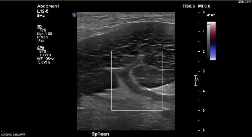
On abdominal ultrasound the spleen was hypoechoic, lacy and lacked bloodflow. This was confirmatory for splenic torsion.
The patient underwent splenectomy and is doing fine.

Files