07/01/19
Publication Date: 2019-07-01
4 images
Findings
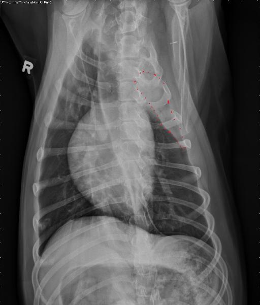
Opposite lateral and VD radiographs of the thorax. An additional VD with intentional rightward obliquity is acquired.
Within the left cranial thorax (best seen on VD views), there is an ill-defined, fusiform soft tissue opaque mass within the region of the left cranial lung lobe cupula. This mass extends cranially from the level of the thoracic inlet, tapering caudally to the level of the left 4th rib. This mass border effaces with the left cranial thoracic wall with a broad-based margin and border effaces with the cranial mediastinum. On the oblique VD view, there is lobar sign between the cranial and caudal sub-segments of the left cranial lung lobe, with a faint air bronchogram seen at the 3rd left intercostal space. At the level of this mass, there are no rib abnormalities and no evidence of extension of the mass laterally beyond the thoracic wall. The mass is not clearly identified on the lateral views. Only on the left lateral view, ventral to the carina, there is a small, mineral opaque, nodule, approximately 1.6cm in diameter.
The cardiovascular structures are normal. The included cranial abdominal structures are normal.
Diagnosis
The described mass is suspected to be origination from the left cranial lung lobe, cranial sub-segment, and may represent a neoplastic process, such as pulmonary carcinoma. An extra-pleural mass, such as sarcoma, cannot be ruled out.
Thoracic ultrasound with fine needle aspiration and/or thoracic computed tomography could be performed for further evaluation.
Discussion
FNAs were performed with ultrasound guidance and were most consistent with a pulmonary carcinoma.

Files