Case of the week 4.22.19
Publication Date: 2019-04-22
History
14 year old male castrated cat. Azotemia. Bronchial disease. HCM. History of nasal lymphoma treated with radiation therapy in 2017
3 images
Findings
Opposite lateral and VD radiographs of the abdomen.
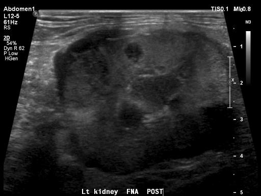
There is moderate soft tissue/fluid opaque streaking within the retroperitoneal space, and focally surrounding the left kidney. The left kidney is markedly enlarged with irregular to lobular margination. There are multiple mineral foci within the plane of the left kidney. The right kidney is small with smooth margination. The urinary bladder is moderately distended and homogenously soft tissue opaque.
The liver, gastrointestinal tract and spleen are normal. There is a large soft tissue opaque nodule within the right caudal lung lobe. Additionally, there is collapse of the right middle lung lobe present on the left lateral view.
There is marked narrowing to almost complete collapse of the L7-S1 intervertebral disc space, with adjacent sclerosis and ventral spondylosis deformans. There is incidental bilateral mild to moderate coxofemoral degenerative joint disease.
Diagnosis
- Left renomegaly and retroperitoneal effusion are concerning for neoplasia (such as renal carcinoma or spread of the previously diagnosed lymphoma)
- Large pulmonary nodule in the right caudal lung lobe again may represent either primary or metastatic neoplasia, such as carcinoma.
- Incidental lumbosacral intervertebral disc disease and coxofemoral degenerative joint disease.
Discussion

FNAs of the left kidney were performed with ultrasound guidance. Results were consistent for lymphoma.
Files