Case of the week 4.8.19
Publication Date: 2019-04-08
History
9 month old female Welsh Corgi. Chronic hypoprotenemia and hypoalbuminemia. Intermittent diarrhea. Low cobalamin.
3 images
Findings
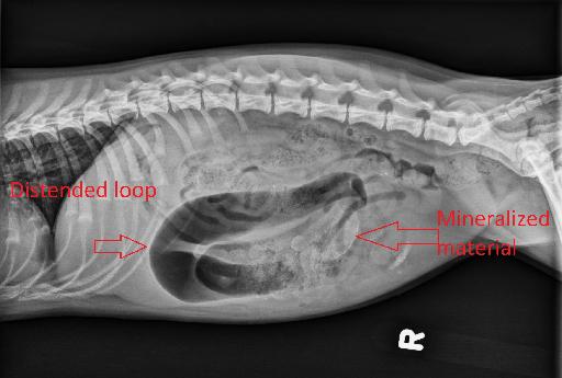
Opposite lateral and VD radiographs of the abdomen.
There is generalized, mild, decreased serosal detail throughout the abdomen, largely due to visceral crowding.
The stomach contains a small volume of gas. There is a mixed population of small intestines with multiple abnormally dilated segments of small intestine. The largest segment of the dilated small intestine is within the cranial ventral abdomen, and is markedly distended with mostly gas. Additionally, within the ventral and right abdomen, likely confined to the small intestines, there is an accumulation of heterogenous, lobular, faintly mineralized, material.
There is a small intestinal segment within the right caudal ventral abdomen on today’s exam which is normal in diameter but contains curvilinear and stippled mineralized material, similar to prior studies. The colon diffusely contains partially formed fecal material.
The remaining intra-abdominal structures are normal. The included thoracic structures are normal. There are numerous open physes, consistent with the patient’s young age.
Diagnosis
The chronic gastrointestinal changes are most consistent with a chronic partial mechanical obstruction. The material described within the right ventral intestines is suspicious for a foreign body. Surgical explore is recommended.
Discussion
The dog underwent surgical explore which confirmed the mechanical obstruction. Material was removed and appeared to be a mixture of cat's hair and feces.

Files