Case of the week 1.9.2024
Publication Date: 2019-03-11
History
2 year old labrador retriever, male castrated.
3 images
Quiz
- The major finding(s) affecting this patient are centered on:The respiratory system
No :-(The cardiovascular system
No :-(The musculoskeletal system
Yes ! :-)The abdominal organs
No :-(
Findings
Opposite lateral and VD projections of the thorax are available for interpretation.
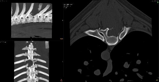
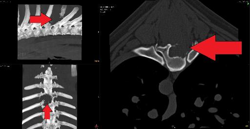
There is lysis of the ventral one half of the spinous process of T7 as well as lysis of the lamina and pedicles of the T7 vertebra. There is the impression of an ill-defined soft tissue opaque mass in the expected region of the ventral one half of the T7 spinous process. Multifocal regions of moth-eaten lysis are noted within the remainder of the spinous process of T7.
The cardiovascular structures are normal. The pulmonary parenchyma, pleural space and mediastinum are normal.
Pinpoint to fine linear mineral opaque foci are seen within the stomach, which also contains mixed soft tissue and gas opaque granular material. The remaining included abdominal structures are normal.
Diagnosis
Monostotic aggressive bone lesion affecting the lamina, pedicles and ventral half of the spinous process of T7. Differential diagnoses include a primary bone tumor (such as fibrosarcoma, chondrosarcoma, or osteosarcoma) or round cell neoplasia (such as lymphoma or plasmacytoma). Potential extension into the vertebral canal and associated spinal cord compression cannot be determined radiographically.
Discussion
CT was performed and the mass was deemed to be non-surgically resectable. The owner elected for human euthanasia.


The dog was initially presented for progressive paraplegia for the last 4 months
Files