Case of the week 3.4.19
Publication Date: 2019-03-04
History
8 year old male intact plott hound. Inappetence, weight loss.
4 images
Findings
Opposite lateral and VD radiographs of the abdomen.
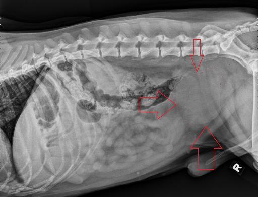
There is fluid streaking of the caudal ventral peritoneal space. Within the caudal ventral abdomen, cranial to the pelvic inlet, and ventral to the colon, there is a large, round, soft tissue opaque mass. This mass is approximately 10 cm in length and 7 cm in diameter, and is resulting in marked dorsal displacement and compression of the caudal descending colon. At the cranial aspect of this mass, superimposed with the small intestines, there is a smaller round to ovoid soft tissue opaque mass (4.5cm x 3.5cm).
The liver is small and there is cranial deviation of the gastric axis. The spleen is normal. The stomach and small intestines are normal. Other than its caudal narrowing and compression, the colon is normal.
Diagnosis
- The large mass of the caudoventral abdomen is suspected to represent a markedly enlarged prostate which may be due to prostatic abscess, prostatitis, prostatic cyst, para-prostatic cyst or neoplasia (such as carcinoma). There is suspect adjacent effusion. The smaller described mass is suspected to represent a cranially displaced and small urinary bladder.
- Microhepatia may be due to a chronic hepatopathy, or represent normal variant.
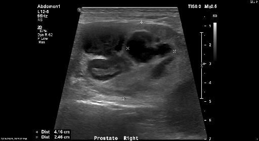
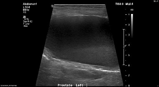
Ultrasound and annotated radiographs
On ultrasound it was noted that there was marked cystic prostatomegaly suggestive of benign prostatic hyperplasia with suspected infection of the large cyst. Prostatitis with abscess formation is also considered. Neoplasia (such as carcinoma) is considered much less likely, yet cannot be entirely excluded.


Diagnosis
The sampling of the fluid were consistent with prostatitis. Staphylococcus Schleiferi was cultured. The dog was discharged on antibiotics and pain management.
Files