Case of the week 2.25.19
Publication Date: 2019-02-22
History
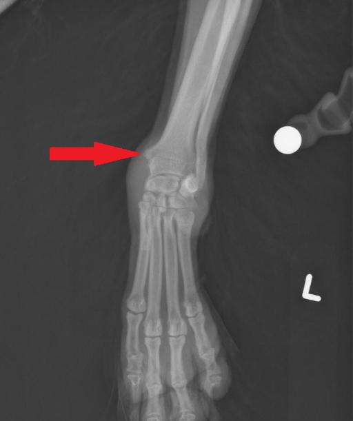
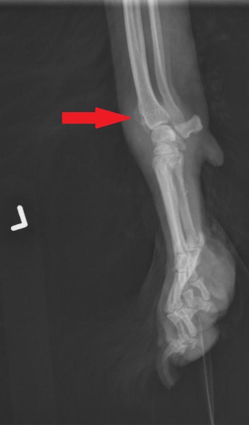
9 year old Cocker Spaniel male castrated. Intermittent lameness left front for the last 5 months.
4 images
Findings
Lateral, dorsopalmar, DMPLO and DLPMO radiographs of the left distal antebrachium, carpus and manus.
There is smooth to curvilinear osseous proliferation at the level of the distal radial groove for the abductor pollicis longus tendon.
There is mild to moderate extracapsular soft tissue swelling medial to the antebrachiocarpal joint. There is also mild intracapsular soft tissue swelling of the antebrachiocarpal and intercarpal joints.
Diagnosis
The changes at the distomedial aspect of the radius are consistent with tenosynovitis of the abductor pollicis longus.
Concurrent suspect carpal joint effusion may be related; however, a separate intra-articular process (such as immune mediate [poly]arthropathy) cannot entirely be ruled out
Discussion
https://onlinelibrary.wiley.com/doi/full/10.1111/j.1740-8261.2011.01886.x
RADIOGRAPHIC AND ULTRASONOGRAPHIC DIAGNOSIS OF STENOSING TENOSYNOVITIS OF THE ABDUCTOR POLLICIS LONGUS MUSCLE IN DOGS Katharina M. Hittmair Veronika Groessl Elisabeth Mayrhofer
Stenosing tenosynovitis of the abductor pollicis longus muscle causes chronic front limb lameness in dogs. The lesion, similar to de Quervain's tenosynovitis in people, is caused by repetitive movements of the carpus. Thirty dogs with front limb lameness, painful carpal flexion, and a firm soft tissue swelling medial to the carpus were examined prospectively. Seven dogs had bilateral abductor pollicis longus tenosynovitis. Radiographs of the carpus were characterized by a deeper radiolucent medial radial sulcus and bony proliferations medial and slightly cranial to the distal radius, resulting in stenosis of the tendon sheath and subsequent tendinitis. Ultrasonographic examination of the firm soft tissue swelling medial to the carpus was characterized by an irregular hypoechoic abductor pollicis longus tendon or tendinitis in 22 of 37 dogs. Nineteen of 37 abductor pollicis longus tendon sheaths were fluid‐filled and all tendon sheaths were thickened, more hyperechoic, with small hyperechoic mineralizations embedded in the connective tissue of the abductor pollicis longus tendon sheath in 25 dogs. Enthesopathy of the abductor pollicis longus tendon was identified in seven dogs. While radiographs of stenosing tenosynovitis of the abductor pollicis longus are helpful in visualizing the deep radial sulcus and osteophytes medial to the distal radius, ultrasonography is useful to distinguish between lesions of the tendon or tendon sheath and to determine thickness and fluid content of the abductor pollicis longus tendon sheath.
In the same article, the treatment part is described as follow: "In dogs,acute abductor pollicis longus tenosynovitis is treated with local methylpredinosolone injections medial to the distal radius and carpus and the area is massaged.After immobilization, this treatment should be repeated. In chronic disease, surgical intervention is required with debridement of the tendon sheath or resection of osteophytes. Tenotomy of the abductor pollicis longus tendon is also performed."
Annoted radiographs


Files