Case of the week 2.18.19
Publication Date: 2019-02-07
History
10 year old mixed breed dog. Anemia, thrombocytopenia, hemorrhagic abdominal fluid
3 images
Findings
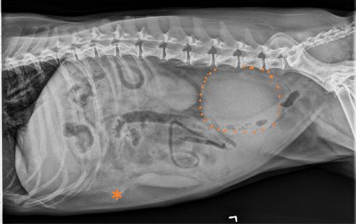
Opposite lateral and VD projections of the abdomen are available for interpretation.
There is mild fluid streaking of the peritoneal fat.
A large, round, well-defined, soft tissue opaque mass is noted in the right caudal retroperitoneal space, ventral to the L4-L7 vertebrae, and is associated with focal ventral deviation of the descending colon and compression of the urinary bladder.
The liver, spleen and kidneys are normal. An elongated, ill-defined, soft tissue opacity is noted in the inguinal region on the lateral views. The gastrointestinal tract is normal aside from the previously described focal ventral deviation of the descending colon.
Diagnosis
Peritoneal effusion is consistent with the reported hemoabdomen.
Large retroperitoneal soft tissue opaque mass. This is most likely neoplastic in origin. This could be non-organ associated and represent an hemangiosaracom in the light of the clinically reported hemoabdomen. Alternatively sublumbar lymphadenomegaly cannot be ruled out. Abdominal ultrasound is recommended for further evaluation.
Additional images


Discussion
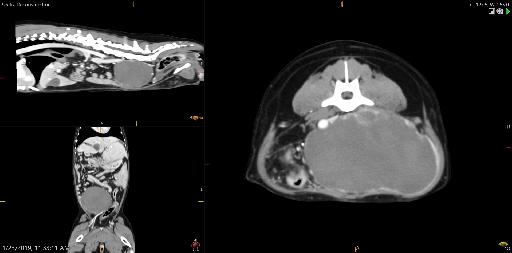
CT was performed. However further treatment was not pursued
The mass was most consistent with a retroperitoneal hemangiosarcoma. Tissue sampling was not obtained.
Files