Case of the week 1.28.19
Publication Date: 2019-01-28
History
12 year old mixed breed dog. Historically lethargic, weak and ataxic
4 images
Quiz
- Based on those images there is:A large splenic mass
Stones in the stomach resulting in gastric outflow obstruction
Stones yes, obstruction noAn enlarged prostate
This a female :)An aggressive bone tumor
no :-)
Additional images


Findings
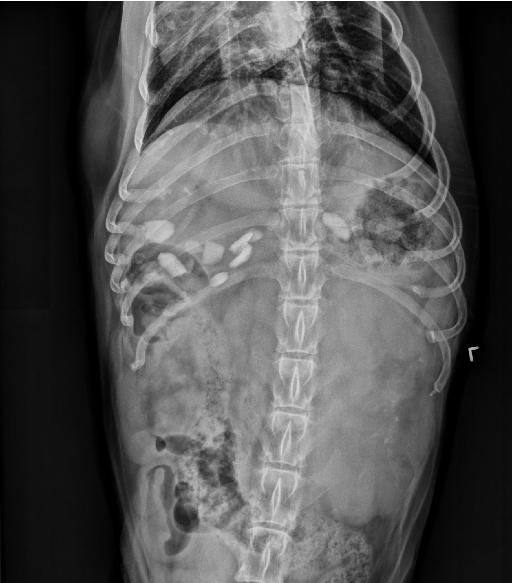
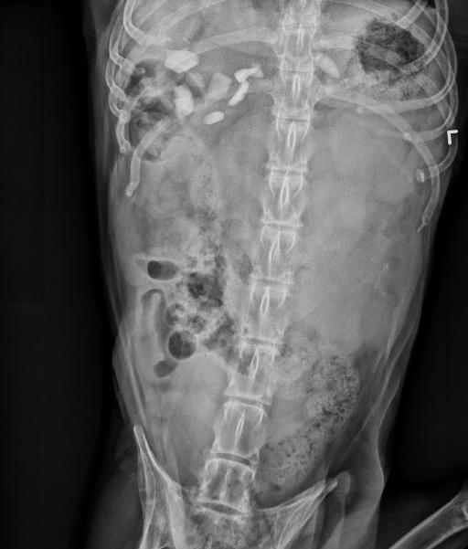
Orthogonal radiographs of the abdomen are available for interpretation.
There is mild decreased serosal detail due to visceral crowding.
Within the left cranial mid abdomen, there is a very large, mostly soft tissue opaque mass with lobular margination and a small region of stippled mineralization. The mass is resulting in marked rightward displacement of the intestines, including the descending colon.
There are multiple mineral opaque angular structures within the stomach. The liver is diffusely moderately enlarged. The splenic head is not clearly identified and the tip of the splenic tail is caudally displaced on the lateral views. The margins of the kidneys are mostly obscured. The urinary bladder is markedly distended and homogenously soft tissue opaque, resulting in mild cranial displacement of the intestines.
There is diffuse osteopenia.
Diagnosis
Large left cranial abdominal mass is suspected to be of splenic or alternatively left kidney etiology, and likely represents neoplasia of malignant (e.g. hemangiosarcoma, carcinoma, other sarcoma) or benign (e.g. hematoma, hemangioma) etiology.
Hepatomegaly is non-specific and could represent benign or malignant etiologies.
Multiple mineral gastric foreign bodies are non-obstructive and likely incidental.
There is no radiographic evidence of pulmonary metastasis. The pulmonary pattern is most likely age related.
Discussion
This case illustrates the importance of acquiring orthogonal radiographs always.
The patient was euthanized and necropsy was not performed.