Case of the week 1.21.19
Publication Date: 2019-01-21
History
13 year old male castrated Basenji. 2 weeks history of inappetence. Left hindlimb lameness for the last two weeks.
3 images
Additional images
a
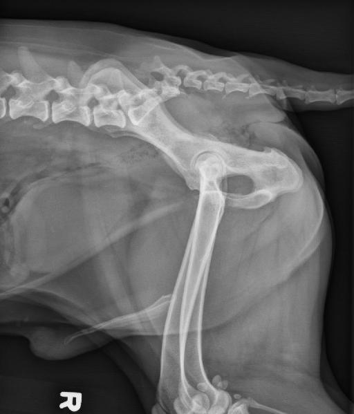
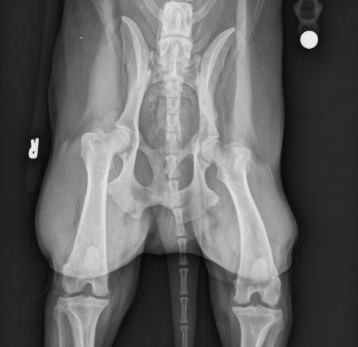
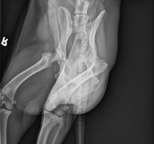
Findings
Orthogonal radiographs of the abdomen are available for interpretation.
The serosal detail is within normal limits.
The liver is mildly small. The caudal pole of the left kidney is smaller than its cranial pole and mildly irregular. There are small pinpoint and linear mineral opacities superimposed with the right mid abdomen. There is an ill-defined soft tissue opaque mass within the pelvic canal displacing the rectum ventrally. The remaining abdominal organs are within normal limits.
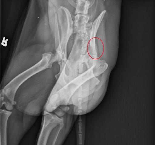
There is a chronic healed malunion fracture of the caudal sacrum and first caudal vertebra. The caudal part of the sacrum and first caudal vertebra are displaced dorsally and cranially and slightly to the right.
There is an ill-defined soft tissue opaque mass within the pelvic canal displacing the rectum ventrally. There is geographic and moth-eaten lysis of the body of the left ilium and of the acetabulum, with lysis of the lateral cortex of the acetabulum and medial cortex of the iliac body. The transition zone is ill-defined. Smooth new bone formation is visible at the lateral aspect of the body of the left ilium. Irregular new bone formation is visible at the dorsal aspect of the body of the left ilium and of the left acetabulum. There is wasting of the left gluteal muscles.
Annotated images
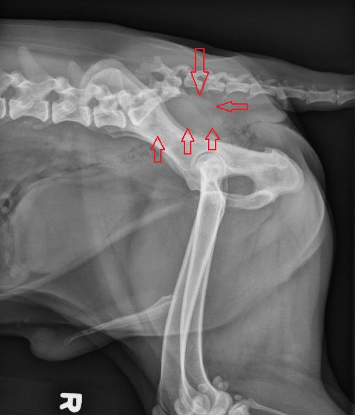
Discussion
1. Aggressive lesion of the left ilium and acetabulum with an intrapelvic soft tissue component is most consistent with a primary bone tumor and less likely a soft tissue neoplasm with bone invasion.
2. Small liver is most likely incidental.
3. The left renal changes are most likely due to chronic renal disease and / or infarcts.
4. The small mineral opacities in the right mid abdomen could represents small calculi in the pancreatic ducts or dystrophic mineralization of the mesenteric vessels.
5. Chronic healed malunion fracture of the sacrum and 1st caudal vertebra.
Diagnosis
Fine needle aspirate of the lytic pelvic lesion and intrapevlic mass were performed.
The pelvic sample was diagnostic for osteosarcoma.
The intrapelvic mass samples were non-diagnostic.
Files