Case of the week 12.17.18
Publication Date: 2018-12-17
History
1 year old female mixed breed dog. Lethargy for the past 5 days. Was in heat2 weeks ago. Might have a portosystemic shunt.
3 images
Findings
Orthogonal radiographs of the abdomen are available for interpretation.
The serosal detail is normal.
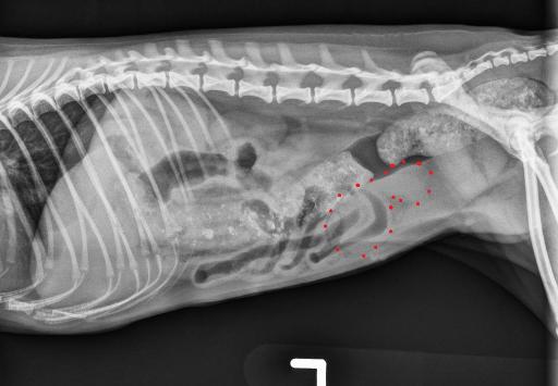
There is a soft tissue opacity, tubular structure superimposed with the cranial and dorsal aspect of the urinary bladder on the lateral projections.
The caudal border of the liver extends to the level of the costal arch and the gastric axis is normal. There is well formed feces in the colon, containing round to rectangular mineral opacities. The small intestines are normal. The spleen and urinary bladder are normal. The kidneys are partially obscured by superimposition of the GI tract, but appear normal.
No abnormalities are identified in the musculoskeletal structures. The caudal thoracic structures are normal.
Diagnosis
The soft tissue structure superimposed with the urinary bladder may represent an enlarged uterus (due to mucometra, hydrometra, early pregnancy, or pyometra), but summation with a fluid-filled small intestinal segment cannot be entirely excluded. An abdominal ultrasound is recommended for further evaluation.
The normal size of the liver makes a portosystemic shunt less likely. Nuclear scintigraphy or a CT scan may be considered for further evaluation.
Discussion
After abdominal ultrasounsd, the patient underwent surgery and confirmed to have a pyometra.
There was no evidence of portosystemic shunt found.

Files