Case of the week 7.30.18
Publication Date: 2018-07-30
History
7 year old mixed breed dog. Male castrated. Suspected intrathoracic mass.
3 images
Findings
Orthogonal radiographs of the thorax are available for interpretation.
There is an ill-defined increased soft tissue opacity at the level of the ascending aorta and aortic arch. This opacity is resulting in mild dorsal deviation of the trachea on the right lateral view and moderate rightward deviation of the trachea on the VD view.
The pulmonary vessels are normal. The pulmonary parenchyma is normal. The remaining intrathoracic structures are normal.
On the right side the right rib is thickened. The included musculoskeletal structures are normal.
A focal round metallic structure is noted superimposed with the falciform fat on the left lateral view.
Diagnosis
- The described soft tissue opacity is most consistent with a heart base tumor, such as the suspect chemodectoma.
- Incidental ballistic projectile of the abdomen.
- Congenitally misshappen second rib on the right side.
Discussion
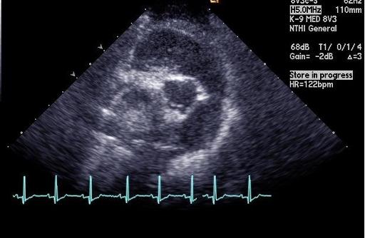
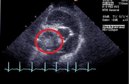
An echocardiogram was performed and confirmed the presence of mass located near the aorta. Differentials include chemodectoma, vs heart base tumor. The patient was started on chemotherapy


Files