Case of the week 7.16.18
Publication Date: 2018-07-16
3 images
Findings
Opposite lateral and VD projections of the thorax are available for interpretation.
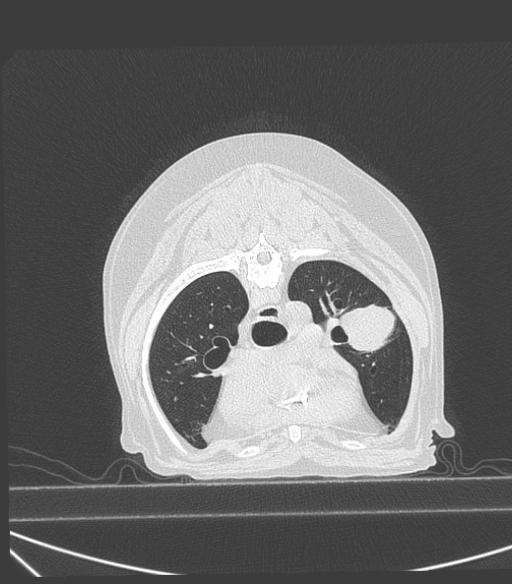
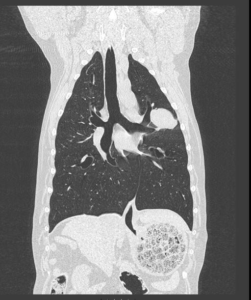
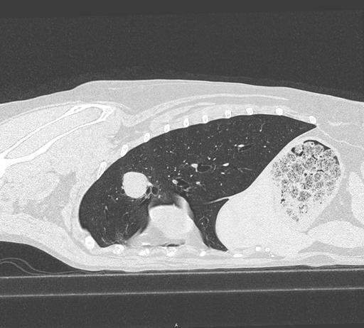
There is a well-defined, ovoid, approximately 4.7 cm x 3.9 cm soft tissue opaque mass in the mid aspect of the cranial subsegment of the left cranial lung lobe. There is the impression of a small, approximately 1.4 cm, ill-defined, round soft tissue opacity in the plane of the right cranial lung lobe, only seen on the left lateral view, superimposed with the fourth ribs. There is an unchanged mild diffuse bronchial and unstructured interstitial pattern as well as persistent faint linear mineralization of the bronchial walls.
The cardiovascular structures remain normal. There is incidental aortic bulb mineralization.
There is marked bilateral shoulder osseous remodeling and periarticular new bone formation. There is narrowing of the T4-T5 intervertebral disc space with sclerosis of the adjacent endplates and spondylosis deformans at this level. There is multifocal spondylosis deformans at the level of multiple other thoracic intervertebral disc spaces.
Diagnosis
Left cranial lobar pulmonary mass is most consistent with a primary pulmonary tumor (such as carcinoma) although pulmonary metastatic disease cannot be entirely ruled out. Suspect additional pulmonary nodule (right cranial lung lobe) is concerning for pulmonary metastatic disease. Ultrasound could be performed to assess the feasibility of ultrasound-guided fine-needle aspiration.
Moderate bilateral shoulder degenerative joint disease.
T4-T5 degenerative intervertebral disc disease.
Discussion
CT was performed for presurgical planning and for evaluation of potential pulmonary metastases. None were found.


The mass was removed surgically and the patient recovered well.
Histopathology was consistent with: LEPIDIC PULMONARY ADENOCARCINOMA
Files