Case of the week 6.11.18
Publication Date: 2018-06-08
History
3 year old female Pomeranian, missing for 2 weeks. Multiple puncture wounds. Unable to use back legs.
6 images
Findings
Opposite lateral and VD projections of the thorax acquired by the emergency service are available for interpretation.
The cardiovascular structures are normal. The pulmonary parenchyma, pleural space and mediastinum are normal. The included musculoskeletal structures are normal.
Opposite lateral and VD projections of the abdomen acquired by the emergency service are available for interpretation.
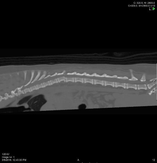
There is ventral subluxation of T13 with regard to T12 resulting in a step in the vertebral canal at this level. There is variation in the degree of subluxation between both lateral views. On the VD view, there is malalignmnet of the T12 and T13 spinous processes, indicative of mild leftward subluxation of T13. There is a transverse fracture of the left 13th rib as well as the impression of a transverse fracture of the left 10th rib.
Numerous variably sized and shaped gas opacities are seen in the subcutaneous tissues of the left caudal abdomen, extending from the dorsum to the ventral abdomen. Numerous punctate to small mineral opaque debris are noted along the skin of the caudodorsal abdomen and perineum.
The serosal margin detail is normal. The liver, spleen, and kidneys are normal. The urinary bladder is small and is of uniform soft tissue opacity. The stomach contains a large amount of mixed soft tissue and gas opaque granular material, consistent with ingesta. The small intestine is normal in size and distribution. The colon contains a small amount of mineral opaque granular fecal material.
Diagnosis
- Ventral and left lateral subluxation of T13 with regard to T12, varying in severity between both lateral views, highly concerning for vertebral instability and associated spinal cord compression.
- Transverse fractures of the left 13th and likely of the left 10th ribs.
- Subcutaneous emphysema along the caudal abdomen and perineum is likely secondary to the reported wounds at these levels.
Discussion
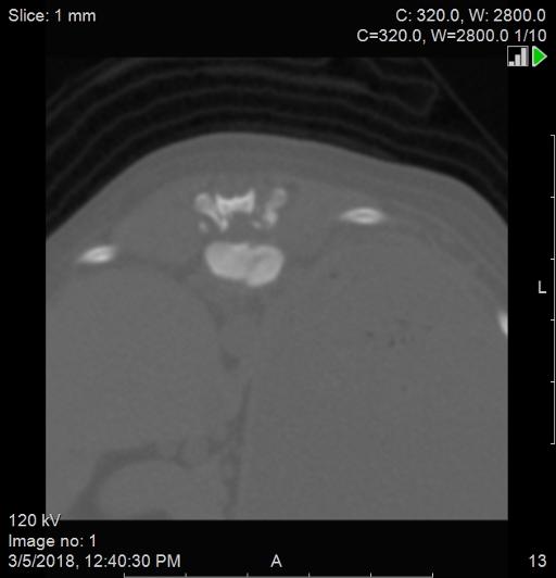
Sagittal and transverse images of the CT illustrate further the radiographic changes. Additionally fractures of T12 endplate, articular facets of T12-T13, and 10th and 13th ribs were also noted.


Outcome
The patient underwent surgical stabilization and is currently doing well.
Files