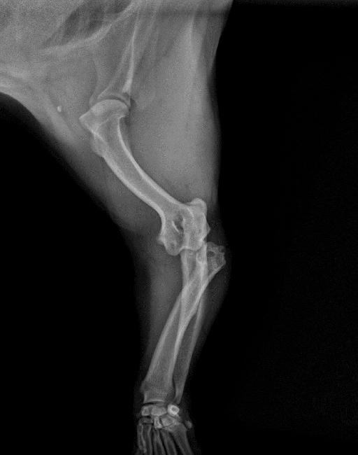
Case of the week 5.14.18
Publication Date: 2018-05-14
History
6 year old American Bulldog. Hit by car 5 days before.
3 images
Findings
Opposite lateral and VD projections of the thorax are available for interpretation.
The cardiovascular structures are normal. There is widening of the cranial mediastinum on the VD view associated with increased fat opacity within the cranial mediastinum on the lateral views. The pulmonary parenchyma is normal. There is mild fluid dilation of the caudal intrathoracic esophagus on the left lateral view.
There are numerous vertebral abnormalities, consistent with the breed. There is mild multifocal thoracolumbar spondylosis deformans. The included abdominal structures are normal.
Files
