Case of the week 4.9.18
Publication Date: 2018-04-09
History
8 year old Greyhound. Was running and stopped.
4 images
Findings
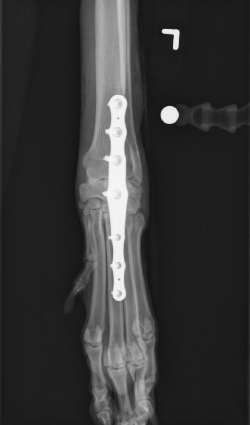
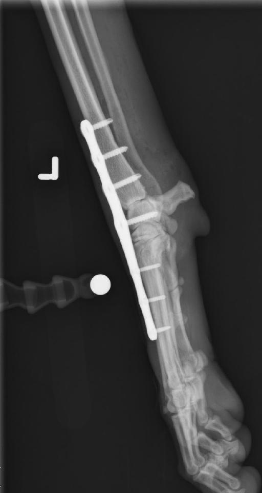
Right carpus, 2 views: Comparison orthogonal views were obtained for surgical planning. No abnormalities are identified. An intravenous catheter is associated with the cranial aspect of the leg.
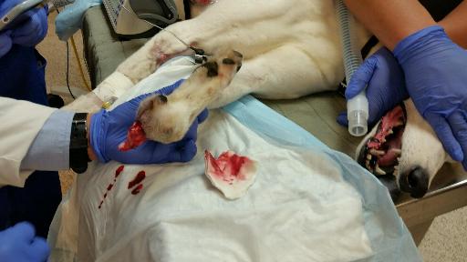
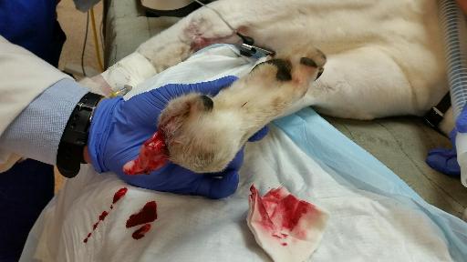
Left carpus, 2 views: There is complete luxation of the antebrachiocarpal joint with carpus and manus flipped 180° and being arranged in a parallel fashion with the antebrachium. There does not appear to be soft tissue coverage of distal antebrachium and proximal carpus.
Diagnosis (GRAPHIC CONTENT)
Complete luxation of the left antebrachiocarpal joint open.


Images, courtesy of Dr. Chambler.
Files

