Case of the week 4.2.18
Publication Date: 2018-04-02
History
8 year old male castrated domestic shorthair. Increased respiratory noises, weight loss
5 images
Findings
Opposite lateral and VD projections of the thorax and neck are available for interpretation.
An approximately 1.5 cm, round, relatively well-defined, soft tissue opaque mass is noted which is confluent with and obscuring the larynx. This mass occupies the entire dorsoventral height of the tracheal lumen at this level.
There is a moderate amount of pericardial and pleural fat. The cardiac silhouette is otherwise normal in size.
There is a mild diffuse bronchial pattern.
There is mild multifocal spondylosis deformans.
The included abdominal structures are normal.
Diagnosis
Laryngeal mass is concerning for malignant neoplasia (such as lymphoma or carcinoma) although a benign process (such as a laryngeal cyst) cannot be entirely ruled out. Ultrasound- or endoscopy-guided tissue sampling of this lesion could be performed for further evaluation.
Mild bronchial pattern may represent chronic lower airway disease, likely of allergic (such as feline asthma) etiology although may also represent a variation of normal for this patient. There is no radiographic evidence of pulmonary metastatic disease.
Ultrasound

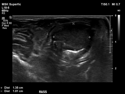
Occupying a majority (~90%) of the lumen of the larynx/cranial trachea, there is a homogenously hypoechoic round mass (1.35cm (W) x 1.0cm (H)).
Fine needle aspirates of the mass were obtained without any complications.
Cytology
Cytology came back as unconclusive as the cells obtained represented a mixed population with evidence of inflammation.
Upon owner's request the patient was discharged with steroids.
Files